
History
The Polish radiology was born in first days of January 1896 in Kraków where the first radiological experiments were performed by Professor Karol Olszewski at the Jagiellonian University. Scientists and physicians in Kraków (mainly connected to Jagiellonian University) are among the pioneers of radiology in Poland or even in the world.
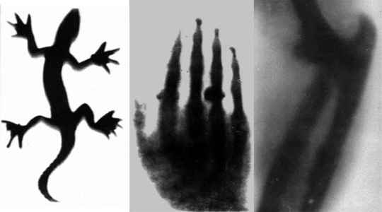
The first Polish X-Ray images (Kraków, January-February 1896)
The Chair of Radiology in Kraków was established on October 15, 1951 as a unit of the Faculty of Medicine of the Medical University of Nicolaus Copernicus (since 1993 Collegium Medicum of the Jagiellonian University). From the very beginning, the headquarter of the Chair was the 17th-century building of the former monastery and then the hospital, at 19 Kopernika St. From the early 1920s it also housed the Department of Radiology of the University Hospital. From January 1, 2023, the headquarter of the Chair was moved to a historic building (1910), the former villa of Professor Piltz (3 Botaniczna St.).
Heads of the Chair of Radiology:
1951-1956 ass. prof. Julian Chudyk – head
1956-1957 dr. Elżbieta Jorasz – acting head
1957-1973 prof. Stanisław Januszkiewicz – head
1973-1975 dr. Halina Naturska-Targosz – acting head
1975-1987 prof. Olgierd Billewicz – head
1987-1998 prof. Józef Kusmiderski – head
1998-2022 prof. Andrzej Urbanik – head
2022- prof. Tadeusz J. Popiela – head
The discovery of X-rays by Wilhelm Konrad Roentgen (on November 8th, 1895) was one of the most important milestones in development of medicine.
The first mention of this discovery in the Polish territory (at that time Poland was partitioned into German, Russian, and Austrian zones; Kraków was at that time in the Austrian zone) appeared in Kraków’s daily Czas of January 8th, 1896. It started with the words “sensational discovery” and the authors referred enthusiastically to the first information about Roentgen’s discovery published just a few days earlier in Vienna’s daily Die Presse by Ernst Lechet on January 5th, 1896. In a short article in Czas there was general information on how the rays were generated, their basic physical properties and hypothetical application. Almost immediately it was noticed that “although this looks like a joke, it is taken seriously by reputable circles”. A few days later, on January 12th, Czas published many more details of the discovery and the technique of using the new rays. It is significant that the publication in Kraków about the discovery of X-rays was one of the first in the world. For comparison, the analogical news were presented in Daily Chronicle on January 6th, The London Standard on January 7th, Berliner Tageblatt on January 8th, Electrical Engineer on January 8th, Electrician on January 10th, Le Matin on January 13th, The New York Times on January 16th.
On the basis of these information, Karol Olszewski, a professor of chemistry at the Jagiellonian University (who was the first in the world, together with Zygmunt Wróblewski, to liquefy air) repeated Roentgen’s experiment. He acquired the equipment necessary for producing X-rays and soon films were taken successfully of different items including a human hand. A report of his experiments appeared in Czas on January 21st, 1896.
From the preserved reports of the experiment that were carried out written by Dr. Tadeusz Estreicher and Dr. Drozdowski (professor’s Ostrowski’s assistant) we learn that in the experiment by Prof. Olszewski air was pumped out from Plucker’s, instead of Crooker’s, tube using Toepler’s pump, and that current was generated by an inducer.
“A photographic plate inside a closed cassette on which a massive wooden box with platinum and brass loads were put, was inserted inside a bag made of thick back cloth, and the whole package was exposed to rays produced by the equipment mentioned. After a long (nearly two hours) exposition Prof. Olszewski developed an image on the plate, which in spite of being faint, did occur. A picture of the bronze load in the shape of a lizard was equally good, also across the wooden bar of the photographic cassette….”
The picture of the bronze load in the shape of a lizard and the picture of Dr. Tadeusz Estreicher’s hand with rings taken at the same time have been preserved. Dr. Drozdowski’s comment on the picture of the hand was as follows:
“Olszewski took this Roentgen picture immediately after the discovery of the rays and just a few days after the announcement of the fact by daily newspapers. He constructed a Roentgen’s tube, of course a very primitive one, that was powered by electric current from a small inductor that was available at the moment, and experimented a couple of hours. Air was being pumped during the experiment and the vacuum was improved by Toepler’s pump…”
According to the documents that have been preserved so far it follows that in his laboratory Prof. Olszewski took successfully a number of pictures using X-rays, while the first radiograms were taken just after the 6th of January. This allows us to assume that these were the first Roentgen pictures taken in Poland. Most certainly there was the picture of the metal paper load among them, the first Polish radiogram. On the other side of the picture there was the following inscription: “First Roentgen film made in Poland, Cracow by Prof. Olszewski”.
At the beginning of February 1896, a professor of surgery, Alfred Obaliński made a decision to use X- rays in practice. His decision followed the admission of a patient with traumatic swollen elbow to his Department of Surgery. In order to find the cause of the symptoms an X-ray examination was made by Professor Olszewski, his assistant Dr. Estreicher, and Dr. Siedlecki. The test radiogram of the elbow joint of a healthy patient was taken. Having found out that in the order to obtain a radiogram two and half hours’ exposure was necessary, the first film of a patient with suspicion of the luxation of the elbow joint was taken. The X-ray examination confirmed the suspicion.
Thus, professor Olszewski was the first Pole to successfully obtain radiograms which preserved until our times and in this way he set the origin of the Polish radiology. A number of X-rayed images were made at the Chemistry Department of the Jagiellonian University on January 6th – 21st, 1896. This fact was reported in “Czas” of 21st January 1896. The first medical report of the first clinical X-ray examination made in Cracow was published in Przegląd Lekarski (publication of Medical Societies – Cracow and Galicia) No 95 (22nd February 1896).
The first Polish scientific radiological article “Diagnostic application of X-rays” was written by Professor Obaliński.
Soon the decision was made by the medical society to introduce X-ray examinations to the clinical practice. The first radiological laboratory was opened in February 1896 (it was the first University X-ray laboratory in Poland) – in the building of the Medical Clinic, in Kopernika Street No. 7 (nowadays housing the Department of Biochemistry of the Jagiellonian University Medical College). It was Dr. Walery Jaworski, subsequently a Professor of the Medical Clinic, who organized this laboratory. He was one of the first physicians all over the world who used X-rays in the clinical practice of internal medicine. On June 16th 1897 at the meeting of the Cracow Medical Society he demonstrated the results of his experiments performed in previous 12 months – for instance a radiological examination of patients with heart enlargement, changes in the aorta and with stones in the gallbladder. This lecture was recognized as the beginning of the Cracovian Radiological School.
Professor Jaworski was strongly interested in stomach diseases. In the handbook “Outline of pathology and therapy of the stomach” he described the Helicobacter pylori bacteria (he named it Vibrio rugula) and suggested that it can cause the diseases of the digestive system. Unfortunately, the book was published only in Polish and the professor wasn’t able to grow such bacteria in a laboratory, so the discovery wasn’t widely known. Only in 2005 Robin Warren and Barry Marshall got Nobel Prize for proving this thesis. Moreover, the research works of Professor Jaworski carried out with the use of contrast media (water with carbon dioxide administrated in to the stomach by a catheter) were of great importance. It was the first this kind of examination in the world. He wrote: “In order to emphasize well the stomach, we will fill it strongly with soda or carbonated powder and the bright spot on the screen corresponding to the stomach will expand and become a kind of a bulging bladder, which is separated from lungs and liver”. As a result it soon became possible to show a picture of an hourglass stomach. The outcomes of all the experiments were published in “Przegląd Lekarski” No 34 and 35 from 21st and 22nd August 1897. Professor Jaworski established also the Historical Museum of the Jagiellonian University Medical College.
The first Radiological Laboratory worked with a primitive equipment constructed by the staff themselves until 1900, when a professional equipment of Reiniger-Halske Company (at present the medical section of Siemens Medical Company) was purchased.
The first Polish handbook of radiology on the “X-rays and their application in diagnostics and therapy” by Dr. M. Nartowski was published in 1900 in Kraków.
In 1913 the new laboratory was established in the St. Lazarus Hospital (it has remained in that place until the 21st century) and it obtained the status of the Department of Radiology. Dr. Barbara Korabczyńska (1881-1949), who devoted her career to that work, was its head.
The works of Professor Jaworski were continued by his former students: Dr Jan Nowaczyński (1885-1925) and Dr Karol Mayer (1882-1956).
Dr. Mayer was the author of the original work “Radiological differential diagnosis of the diseases of the heart and the aorta with the consideration of own methods”, published in 1916, where he included the elements of tomography, unknown at that time.
Although the work of Mayer was unnoticed in the radiological world because of the time and place (the period of World War I, the absence of Polish on the map of Europe), it should be emphasized that he defined the problem properly: “… organs have in fact three dimensions and are arranged in space, however in the radiological image they all are projected on one plane of two dimensions, so as a result the structures which in fact are one outside the other, in the image they are located next to each other or cap each other…” and its solution: “… I remove such difficulties as follows: After the usual preparations for pictures I make fast and small (up to 8 cm) X-ray tube moves back and forth parallel to the longitudinal or transverse axis (according to the case) of the body during the whole exposition. These movements can be easily and completely safety performed using tripods at which the lamp is placed in easy shifting box. In this simple way I achieve a complete disappearance of the shadows which do not belong to the shadow of the heart and blood major, or a partial separation of them, so that what seeing the shape of the individual curves becomes possible…”. Mayer’s discovery was seven years before the presentation of the theoretical principles by A. Boccage and getting his patent in 1921 (this date was taken as the emergence X-ray tomography). Moreover, K. Mayer immediately applied in practice his idea (in so difficult anatomical areas like a chest).
In 1914 Mayer received in Germany a patent (Patentschrift No. 274790) on your own idea of the X-ray lamp provided with two or more anodes and cathodes. He worked under the supervision of Professor Jaworski, and in 1925 he was appointed the Head of the Radiology Department of the Poznań University, and soon became the first Polish Professor of Radiology.
The building of the Department of Neuropsychiatry at the Jagiellonian University was completed in 1914. During the World War I it was a military hospital and only since 1919 there were treated civil patients and the building became a seat of the clinics (it’s head was Professor Jan Piltz). Since 1934 next to the clinic there was a general radiology lab (over 10 years later the polish neuroradiology was born here), which was led by Teofil Bluhbaum (1900-1971).
In 1926 the first polish scientific radiological journal was established – Polski Przegląd Radiologiczny (Polish Radiological Review). One of its founders was the radiologist from Kraków, Dr Henryk Wachtel.
November 6th, 1930 the Kraków Section of the Polish Medical Society of Radiology (the Society was founded in 1925).
In 1945 dr. Stanisława Spettowa (1902-1994) came to Kraków from Lviv (it was a Polish city before the World War II) and on 1st August took over management of the Radiological Laboratory in Department of Neuropsychiatry. Over 26 years Professor Spettowa created in Kraków a neuroradiological center at the highest level, which was a beginning of the polish neuroradiology.
Dr Korabczyńska died in 1949 and left the Department of Radiology as a big diagnostic and therapeutic institution. Then Dr. Julian Chudyk (1894-1956) was the Head of the Radiology Department. He organized the XII Congress of the Polish Medical Society of Radiology in 1949.
In 1951, after the reform of higher medical education, the Faculty of Medicine in Kraków, the oldest one of the Jagiellonian University, was separated and the Medical University was created instead. The Department of Radiology of the University Hospital was founded in March, and the Chair of Radiology on October 15, 1951 with Dr Chudyk as its Head (since 1954 he was an Associate Professor). The Department was modernized as the research-didactic-diagnostic unit. The successor of Professor Chudyk was Dr. Elżbieta Jorasz, the acting head of the Department after his death (1956-57).
Professor Stanisław Januszkiewicz (1903-1979) was the head of the Department and Chair of Radiology between 1957 – 1973. He was the author of the handbook “General radiological diagnostics – student’s handbook”. In 1935 received a patent (no 21878 on September 25th, 1935) on the prototype of submersible iris which is now widely used. In June 1964 Professor Januszkiewicz organized in Kraków the XXII Congress of the Polish Medical Society of Radiology. After he retired in 1973, his duties were taken up by Dr. Halina Naturska-Targosz (1973-1974) as the acting head.
The handbook “Neuroradiology” was published in 1970. It was written by Associate Professor Ryszard Chrzanowski (1930-2022) who was between 1973-74 the Head of the Department of the Neuroradiology in Institute of Neurology in Kraków. His successor was Professor Józef Kuśmiderski (1928-1998).
Since 1975, the Head of the Chair and Department of Radiology was Professor Olgierd Billewicz until he moved to Gdańsk in 1988. During that period new diagnostic techniques: computed tomography (1982), mammography (1983) and ultrasound (1983) were introduced. All these laboratories were the first in South-East Poland. The modern two-plane angiograph was bought in 1977. The radiological interventional procedures were introduced in 1986 by Dr Andrzej Urbanik (1953-).
The XXXI Congress of the Polish Medical Society of Radiology took place in Kraków on June, 1986. Professor Billewicz, the organizer of the Congress, was at that time elected the President of the Main Board of the Society. Dr Andrzej Urbanik, an assistant in the Department, was appointed the Secretary of the Board.
Professor Józef Kuśmiderski became the Head of the Chair and Department of Radiology in 1988. He was at the same time the Head of the Neuroradiology Department (since 1974). Under his supervision, the plan of creating a new seat of the Radiology Department in 50 Kopernika St. was developed and the construction works started (1996).
On May 12th, 1993 The Faculty of Medicine returned to Alma Mater Jagellonica and was a part of Collegium Medicum (Medical College) which was created at the Jagiellonian University.
In 1994 the DSA angiograph was installed in the Department of Neuroradiology and neuroradiological interventional procedures were introduced. First treatments were performed by Dr. Andrzej Urbanik.
The Chair of Radiology cooperated with the Institute of Nuclear Physics in Kraków where in 1985 a group of scientists leaded by Prof. Andrzej Jasiński (Department of Radiospectroscopy) worked on the magnetic resonance (MR) system. The Institute of Nuclear Physics in Kraków is an important institution in field of MR. Two employees – physicists Andrzej Hrynkiewicz and Jacek Hennel received there in 1953 the first MR signal in Poland. They constructed the whole necessary equipment by themselves using even an old former German military radio station.
The 2nd Congress of the Polish Medical Society of Magnetic Resonance was organized by Professor Kuśmiderski in Kraków on September 1996. The Congress celebrated the centenary of the Polish Radiology whose origin has to be traced in Kraków. On that occasion a jubilee exhibition was organized in the Jagiellonian University Museum – “Pioneers of Cracow’s Radiology – one hundred years” (curators: Dr. Ewa Wyka and Dr. Andrzej Urbanik).
In 1998, after Professor Kuśmiderski’s death, his duties were taken over by Prof. Andrzej Urbanik who was appointed the Head of the Chair of Radiology of the Jagiellonian University Medical College and the Head of the Radiology Department of the University Hospital.
In the years 1998-2000 the MR laboratory building was finished and the main building housing the Chair and Radiology Department in 19 Kopernika St. was fully modernized and modern equipment that meets global standards was provided. A very important moment for radiology in Kraków was opening the Magnetic Resonance Laboratory at the Chair of Radiology in 1998. It was equipped with the first-class 1,5T MR system. Not only standard examinations but also a variety of new techniques were introduced and research-scientific studies were made. For the first time in Poland a variety of new MR techniques were used: phosphorus spectroscopy, functional brain imaging, MR evaluation of flow of cerebrospinal fluid, breast biopsy under the control of MRI, complex (containing spectroscopy) prostate imaging.
In 1998 in the Chair and Department of Radiology for the first time in Poland new information technology systems were introduced: RIS (Radiological Information System) and teleradiology as well as a virtual endoscopy option. One year later PACS (Picture Archivisation and Communication System) was launched.
The team of radiographers and physicians leaded by Prof. Andrzej Urbanik finished in 2000 complex radiological research (conventional X-ray and computed tomography) of Egyptian mummies which are in Polish collections. The program was run since 1995. It was one of the first experiments using 3D techniques and virtual endoscopy. A pioneer method of mummy’s face image reconstruction was invented.
In 2001 in the Chair and Department of Radiology there was the first in Poland angiograph with the option of rapid rotation and 3D reconstruction which allows for new opportunities in angiodiagnostics especially in central nervous system.
In 2001 the system for indirect digital radiography was tested and then such system was introduced so that the classic radiography was eliminated.
In 2001/2002 the Rapid Prototyping technique was introduced (first time in Poland) – models of the body parts based on radiological images (mainly CT) can be created.
In 2005 for the first time in Poland the DynaCT option was installed which allows for making images similar to the computed tomography.
The first in Central-East Europe system RheumaLab was launched in Department of Diagnostic Imaging in 2007. It is designed to diagnostics of rheumatologic diseases and contains MR system dedicated to examinations of joints, ultrasonograph as well as a system for imaging fusion and stereotactic puncture.
The Institute of National Remembrance outsourced in 2008 to the Chair of Radiology preparing examination of the exhumed remains of General Władysław Sikorski (he died in unclear circumstances in a plane crash in Gibraltar on July 4th, 1943). X-ray and CT investigations were performed. Based on the image data an extensive report was compiled. The cooperation with the Department of Forensic Medicine was established then. Since 2009 CT examinations made by radiologists precede actual autopsy in some court cases. Later a series of MRI was performed for the same purposes. These examinations were the first in Poland.
Due to the building of Combined Departments of General Surgery, Endoscopic Surgery and Rescue Medicine and Multiorgan Trauma (CUMRiK) at the University Hospital in Kraków, in this unit there was the Department of Diagnostic Imaging created in October 2012 (the head: Ass. prof. Tadeusz J. Popiela)
At the Faculty of Health Sciences at the Jagiellonian University Medical College the Department of Electroradiology was established (2012) under the Institute of Physiotherapy. It was the culmination of many years of efforts of the Chair of Radiology to create at the Medical College a special unit educating radiographers at academic level. The head of the new Department became Ass. prof. Izabela Herman-Sucharska (previously lecturer in the Chair of Radiology).
In 2019, a new University Hospital was launched in Kraków, in the district of Prokocim, at No. 2, Jakubowskiego Street. Most clinics were transferred there. A new Imaging Diagnostic Department with state-of-the-art equipment was also established at the new hospital, with Prof. Tadeusz Popiela appointed as the Head of the Department. A few clinics remained at the former location, and the related services were provided to these by the existing Imaging Diagnostic Department (headed by Prof. Andrzej Urbanik); the laboratories were located in three buildings (at 19 Kopernika St. – main office, 17 Kopernika St., and 50 Kopernika St.). In 2022 Prof. Andrzej Urbanik retired from work. The University Hospital Imaging Diagnostic Department was then reorganised into a single unit, with Prof. Tadeusz Popiela as the Head of the Department. Since 2023 the major part of the Radiology Department is located in the new Hospital University (at 2 Jakubowskiego St.) and the remaining facilities at 50 Kopernika St. Prof. Tadeusz Popiela was also appointed the Head of the Chair of Radiology, whose main office was moved to the historical building at No. 3, Botaniczna St. (Professor Piltz’ villa).